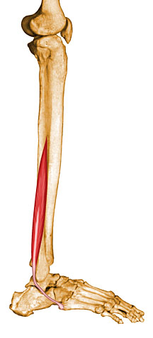
view of the peroneal brevis tendon
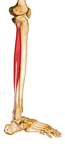
view of the peroneal longus tendon
Peroneal Tendon Injury
Anatomy & Synopsis
The peroneal tendons are two tendons that lie
immediately behind the outside bone of the ankle (the fibula). These two tendons
are responsible for moving the foot outwards in a direction called eversion.
They are important tendons because they balance the ankle and the back of the
foot and prevent the foot from turning inwards repetitively. They are slightly
weaker than the muscles and tendons on the inside of the ankle and are prone to
injury as the ankle turns, rolls or becomes sprained.
|
view of the peroneal brevis tendon |
view of the peroneal longus tendon |
Tears of these tendons do occur. One or both of the tendons can be torn. This
leads to swelling, pain and a sense of instability behind the outside of the
ankle. Occasionally the tendons can be injured in either a fall or an athletic
injury. They pop out of the supporting ligaments that hold them in place and
dislocate. Once this occurs, recurrent dislocation and tearing of the tendons is
inevitable.
If the tendons dislocate acutely in an injury, they need to be repaired to
prevent future tearing of the tendons.
Diagnosis And Treatment
The diagnosis of peroneal tendon injury is made through careful examination and
palpation by the orthopedic surgeon. An MRI may be required to more clearly
document the extent of the tear. Once a tear is diagnosed, surgery is necessary.
The tendons can be repaired by stitching. If they are severely torn, they need
to be replaced with new tendon tissue. Sometimes this tendon tissue can be
obtained from the same leg. At other times allograft tendons are used. These
come from the cadaver bank. The allograft tendons are safe to use, have no
immunogenic properties and are not rejected. This procedure has been
successfully performed on professional athletes who have returned to national
level competition.