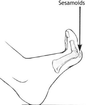
the sesamoids act like pulleys.
a view looking in from the anterior of the foot. note that this particular foot has sesamoids under each toe!
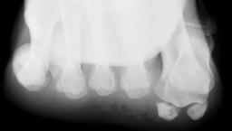
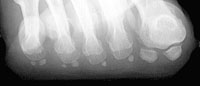
typical sesamoid x-ray
Sesmoiditis, Biparte Sesamoids
Source: American Academy of Orthopedic Surgeons
(revised 03/07/2006)
Synopsis
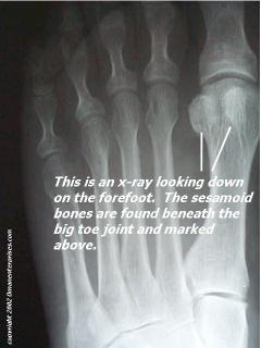
Most bones in the human body are connected to each other at joints. But there are a few bones that are not connected to any other bone. Instead, they are connected only to tendons or are embedded in muscle. These are the sesamoids. The kneecap (patella) is the largest sesamoid. Two other very small sesamoids (about the size of a kernel of corn) are found in the underside of the forefoot near the great toe, one on the outer side of the foot (lateral) and the other closer to the middle of the foot (Medial). these sesamoids are embedded in the flexor hallucis brevis tendon, one of several tendons that exert pressure from the big toe against the ground and help initiate the act of walking.It is not uncommon for people to have more then two sesamoids on the great toe as well. this condition is called Biparte sesamoids and can easily be misdiagnosed as a fracture of one of the medial or lateral sesamoids, when infact it is a third sesamoid in its own right. Because the edges of a bipartite medial sesamoid are generally smooth, and the edges of a fractured sesamoid are generally jagged, an X-ray is useful in making an appropriate diagnosis. Your physician may also request X-rays of the other foot to compare the bone structure.
|
the sesamoids act like pulleys. |
a view looking in from the anterior of the foot. note that this particular foot has sesamoids under each toe!
typical sesamoid x-ray |
Sesamoids act like pulleys. They provide a smooth surface over which the tendons slide, thus increasing the ability of the tendons to transmit muscle forces. The sesamoids in the forefoot also assist with weight bearing and help elevate the bones of the great toe. Like other bones, sesamoids can break (fracture). Additionally, the tendons surrounding the sesamoids can become irritated or inflamed. This is called sesamoiditis and is a form of tendonitis. It is common among ballet dancers, runners and baseball catchers.
Signs and symptoms
Examination and diagnosis
During the examination, the physician will look for tenderness at the sesamoid bones. Your doctor may manipulate the bone slightly or ask you to bend and straighten the toe. He or she may also bend the great toe up toward the top of the foot to see if the pain intensifies. Your physician will request X-rays of the forefoot to ensure a proper diagnosis. If the X-rays appear normal, the physician may request a bone scan.
Treatment
Treatment is generally non-operative. However, if conservative measures fail, your physician may recommend surgery to remove the sesamoid bone.
- Stop the activity causing the pain.
- Take aspirin or ibuprofen to relieve the pain.
- Rest and ice the sole of your feet. Do not apply ice directly to the skin, but use an ice pack or wrap the ice in a towel.
- Wear soft-soled, low-heeled shoes. Stiff-soled shoes like clogs may also be comfortable.
- Use a felt cushioning pad to relieve stress.
- Return to activity gradually, and continue to wear a cushioning pad of dense foam rubber under the sesamoids to support them. Avoid activities that put your weight on the balls of the feet.
- Tape the great toe so that it remains bent slightly downward (plantar flexion).
- Your doctor may recommend an injection of a steroid medication to reduce swelling.
- If symptoms persist, you may need to wear a removable short leg fracture brace for 4 to 6 weeks.
- You will need to wear a stiff-soled shoe or a short, leg-fracture brace.
- Your physician may tape the joint to limit movement of the great toe.
- You may have to wear a J-shaped pad around the area of the sesamoid to relieve pressure as the fracture heals.
- Pain relievers such as aspirin or ibuprofen may be recommended.
- It may take several months for the discomfort to subside.
- Cushioning pads or other orthotic devices are often helpful as the fracture heals.