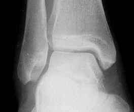
Normal Ankle
Anatomy and
Function
The
ankle works in a systematic way. Movement is only supposed to be in one plane,
in other words, up and down. We call this dorsiflexion and plantarflexion. The
ankle joint is held in place securely by a group of bones that house the main
anklebone (called the talus) inside a box-like effect. On the inside is the
medial malleolus and on the outside the fibula.
|
Normal Ankle |
The inward and outward movements of the back of the foot do not actually occur
in the ankle joint but occur in the joint underneath it called the subtalar
joint. The muscle that pulls the foot inward (inversion) is slightly stronger
than the muscles that pull the foot outward (eversion). When the foot lands in
an awkward manner there is a tendency for the heel to roll inwards and create
stress on the outside ligaments. If this stress is severe then a sprain of the
ankle occurs. A sprain is actually a tear that occurs in the outer supportive
ligaments of the ankle. As these ligaments are stretched, a critical point is
reached beyond which ligaments do not return to their normal elastic function
and a tear of the ligament occurs. Sprains can range from the relatively minor
to those where the ligaments are completely torn and the ankle can be quite
loose.
|
|
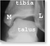
Front
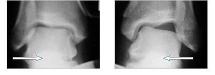
of the ankle. M= medial malleolus, L=lateral malleolus (fibula). The outside of the heel is pushed inwards to stress the joint. In the normal ankle, no tilting of the ankle should occur at all. Note the tilting of the talus in the ankle. |
The acute sprain of the ankle is commonly associated with marked swelling and
bruising on the outer side of the ankle. Rest of the ankle with immobilization
of some sort is critical. The classic treatment for a sprain of the ankle is
what we refer to as the Rice Program. It involves rest, ice, compression and
elevation. This treatment is designed to decrease the inflammation and swelling
of the ankle associated with the sprain. The Rice Program by itself will not
heal the ligaments. In order for the ligaments to heal the ankle needs to be
immobilized with either a cast or a boot. For minor sprains a brace can be
applied to the ankle. Walking is permitted during this recovery process,
allowing the ankle ligaments to heal.
Following this period of initial immobilization, strengthening exercises are
essential to regain the balance of the ankle. It is critical that the tendons
and muscles on the outside of the ankle (the peroneal tendons) are strengthened.
This should be done initially in a supervised exercise program. If the ligaments
have been severely torn, the ability to fine tune the ankle and prevent further
sprains from occurring depends on the strength of the peroneal muscles. As the
ankle turns repeatedly, the peroneal muscles weaken further. This weakens the
ability to prevent recurring sprains. Patients with a high arch or a heel that
is naturally turned in slightly are predisposed to sprains.
As a result of continued rolling, turning or instability of the ankle, the
ability to fine tune the foot on uneven surfaces becomes limited. The ability to
make rapid changes in the position of the foot on the ground surface is called
proprioception. If this ability is diminished, the likelihood of a more severe
ankle sprain occurring is increased. In recurring ankle sprains we call this
chronic recurrent instability of the ankle. The ankle is at risk of developing
other problems. These include bruising of the cartilage of the talus and bone
spurs that develop around the front and sides of the ankle. These are all
precursors of ultimate arthritis of the ankle.
 |
||
|
||
The diagnosis of chronic recurrent instability is made through a careful
examination of the ankle and X-rays that are taken while stress is applied to
the ankle.
 |
||
|
||