Tarsal
Coalition
Synopsis
Tarsal
Coalition is a condition where one or more bones are abnormally joined together
(fused or coalesced) in the back of the foot. Normal development of any joint
requires that the bones on either side of the joint move freely.
A child born with Tarsal coalition has bones that do not develop normally
because the joint and its cartilage lining never fully develop. For this reason,
the two bones that are involved in the coalition are stuck together and the foot
does not move in and out (inversion and eversion) adequately.
As the foot develops, the child and family will notice that the foot is flat and
often getting progressively flatter as the child gets older.
 |
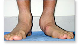 |
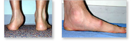
These pictures are of a 16-year-old boy with a tarsal coalition
of one foot. Note how flat the foot is. |
|
Symptoms include aching, soreness and fatigue after
exercise and specific soreness and pain in the back of the foot and heel joint.
When symptoms from tarsal coalition begin, they can be quieted down by
immobilizing the foot in a cast or a boot. This does not treat the underlying
problem of the coalition but it does help temporarily with the symptoms of pain
and soreness. Occasionally, the use of a cast for a few months will be
sufficient as treatment and once the cast is removed the symptoms dissipate.
While the symptoms of the coalition may be improved, the coalition remains, and
the foot continues to be stiff. For this reason, once a coalition is diagnosed,
it is unusual that the use of the cast immobilization is the only form of
treatment.
Diagnosis
and Treatment
The diagnosis
of a tarsal coalition can easily be made by a careful examination of the
movement segments of the back of the foot. This is then confirmed with x-rays. A
CAT scan or an MRI will rarely be needed to confirm the diagnosis.
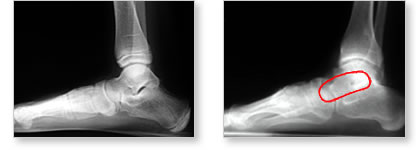 |
| These
are X- Rays looking at the side of the foot. On the foot on the
right, there is no space between the heel bone (calcaneus) and
the ankle bone (talus). The space is taken up by abnormal bone,
called a coalition. The foot on the left is a normal XR where
you can see the space between the calcaneus and the talus, and
the one on the right has the coalition circled in red. |
|
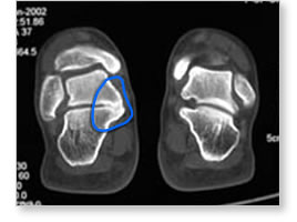 |
| If
it cannot be seen on a plain X-ray, the diagnosis of a tarsal
coalition becomes more obvious using a CAT scan. Note in the
scan here, the normal foot on the right and the bone connection
between the talus and calcaneus on the left. |
|
Depending on how uncomfortable or painful the foot is, treatment can be
initiated with a cast, boot or a brace (STEPS AFO) and then followed with
orthotic arch supports. As noted above, the boot or orthotic support will not
ever be curative because the tarsal coalition and the abnormal bone connection
are still present. In order to completely eliminate symptoms and improve the
movement in the back of the foot, surgery is required. The surgery is designed
to remove the tarsal coalition and improve the inversion and eversion movement
of the foot. Once this is done, most children are able to resume full athletic
activity. There are times when removal of the tarsal coalition cannot completely
correct the deformity. For these children, additional surgery may be necessary,
particularly if the tarsal coalition if associated with a very flat foot. This
is because simply removing the tarsal coalition will not correct the arch of the
foot itself.
The recovery following removal of a tarsal coalition is straightforward. A
removable boot is worn on the foot for six weeks. Walking on the boot can
commence at two weeks during which time, strengthening and various exercises are
done on a regular basis to maintain and improve the movement and strength and
arch of the foot.